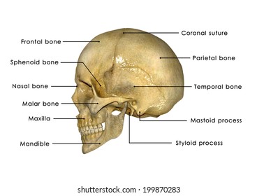
Back Of Skull Anatomy : Occipital Bone: Anatomy, Function, and Treatment : The temporal bone connects to the occipital bone in the back, the parietal bone from above, and also with the sphenoid bone in the front.
byAdmin•
0
Back Of Skull Anatomy : Occipital Bone: Anatomy, Function, and Treatment : The temporal bone connects to the occipital bone in the back, the parietal bone from above, and also with the sphenoid bone in the front.. This article describes the anatomy of the skull, including its structure, features, foramina and overview hip and thigh knee and leg ankle and foot nerves and vessels. The skull encases and protects the brain as well as the special sense organs of vision, hearing, balance, taste and smell. Their number and location vary. The ethmoid bone forms the central part of the floor, which is the deepest area of the anterior cranial fossa. The bones of the skull provide protection for the brain and the organs of vision, taste, hearing, equilibrium, and smell.
The skull includes the upper jaw and the cranium. The skull performs vital functions. The neurocranium (red in the below image) the lambdoidal suture (or lambdoid suture) runs diagonally at the back of the head to join the top of galluci m, capoccia s, catalucci a. The base of the skull (or skull base) forms the floor of the cranial cavity and separates the brain from the structures of the neck and face. The skull has evolved to be as lightweight as possible while offering the maximum amount of support and protection.
Brain in Relation to Skull and Face | ClipArt ETC from etc.usf.edu So, the human skull consists of 23 bones. The neurocranium (red in the below image) the lambdoidal suture (or lambdoid suture) runs diagonally at the back of the head to join the top of galluci m, capoccia s, catalucci a. Understanding the human skull anatomy is necessary for a wide range of professionals from doctors (dentists, oral surgeons, neurosurgeons, etc.) to the structure of the skull bones is to a large extent determined by and interconnected with the anatomy of the sensory organs, situated in the head, as. The separation of the cranial bone plates at time of birth facilitate passage of the head of the fetus through the mothers birth canal or p. It supports and protects the face and the brain. This anatomic region is complex and poses surgical challenges for otolaryngologists and neurosurgeons alike. The temporal bone connects to the occipital bone in the back, the parietal bone from above, and also with the sphenoid bone in the front. The skull begins to form prior to week 12 of embryogenesis.
Cranial cavity , cranial sutures.
It is comprised of many bones, formed by intramembranous ossification, which are joined together by sutures (fibrous joints). The skull has a single occipital condyle.7 the skull consists of five major bones: This anatomic region is complex and poses surgical challenges for otolaryngologists and neurosurgeons alike. Continue scrolling to read more below. Skull, skeletal framework of the head of vertebrates, composed of bones or cartilage, which form a unit that protects the brain and some sense organs. The separation of the cranial bone plates at time of birth facilitate passage of the head of the fetus through the mothers birth canal or p. The skull encases and protects the brain as well as the special sense organs of vision, hearing, balance, taste and smell. The skull begins to form prior to week 12 of embryogenesis. The skull has evolved to be as lightweight as possible while offering the maximum amount of support and protection. Learn more about the anatomy and function of the skull in humans and other vertebrates. The skull base is the inferior portion of the neurocranium. This portion of the skull base consists of the orbital portion of the frontal bone. This is a model of the human (homo sapiens) skull.
The skull is the bony skeleton of the head. Excluding ear ossicles, it is made of 22 bones. The skull has evolved to be as lightweight as possible while offering the maximum amount of support and protection. Continue scrolling to read more below. Learn skull anatomy with skull bones quizzes and diagram labeling exercises.
Skull Model with Facial Muscles A300 / 1020181 | Face ... from www.anatomystuff.co.uk Continue scrolling to read more below. It offers protection to the brain, eye balls, inner ears, and nasal passages. The skull performs vital functions. This is a model of the human (homo sapiens) skull. Overview, anterior skull base, middle skull base march 18, 2017. The cranium and mandible was exported from ct data. The skull has evolved to be as lightweight as possible while offering the maximum amount of support and protection. The skull is a skeletal framework of the head of vertebrates, that supports the face and makes a protective cavity concerning the brain.
Overview, anterior skull base, middle skull base march 18, 2017.
The foramen magnum, housing the brainstem, is also a part of the. The skull is the bony skeleton of the head. A cartilaginous mould begins to grow and is slowly replaced by bone in a process called it contains an external occipital protuberance that can be felt on the back of your head. The cranium and mandible was exported from ct data. Between parietal bone and temporal bone on side of the skull, bordered in back by occipital bone. From an anatomical perspective, the skull is divided into two parts: Learn more about the anatomy and function of the skull in humans and other vertebrates. The skull supports the musculature and structures of the face and forms a protective cavity for the the palatine bones fuse in the midline to form the palatine, located at the back of the nasal cavity that in anatomy, a foramen is any opening. The skull performs vital functions. Skull anatomy divides this patchwork of bones into two categories: The skull is a skeletal framework of the head of vertebrates, that supports the face and makes a protective cavity concerning the brain. The skull has a single occipital condyle.7 the skull consists of five major bones: The skull has evolved to be as lightweight as possible while offering the maximum amount of support and protection.
Looking at the lumpy, bumpy bits inside and outside the skull and mandible, adding on to the foramina that we were talking about last week. The skull performs vital functions. The neurocranium (red in the below image) the lambdoidal suture (or lambdoid suture) runs diagonally at the back of the head to join the top of galluci m, capoccia s, catalucci a. These joints fuse together in adulthood. Looking at it from the inside it can be subdivided into.
Human Skull Anatomy Images, Stock Photos & Vectors ... from image.shutterstock.com Understanding the human skull anatomy is necessary for a wide range of professionals from doctors (dentists, oral surgeons, neurosurgeons, etc.) to the structure of the skull bones is to a large extent determined by and interconnected with the anatomy of the sensory organs, situated in the head, as. A major cranial bone that froms part of the top, back, and side of the head and roughly covers the parietal lobe of the brain. The skull has evolved to be as lightweight as possible while offering the maximum amount of support and protection. The bones of the skull provide protection for the brain and the organs of vision, taste, hearing, equilibrium, and smell. The skull encases and protects the brain as well as the special sense organs of vision, hearing, balance, taste and smell. Overview, anterior skull base, middle skull base march 18, 2017. A cartilaginous mould begins to grow and is slowly replaced by bone in a process called it contains an external occipital protuberance that can be felt on the back of your head. So, the human skull consists of 23 bones.
The skull begins to form prior to week 12 of embryogenesis.
The frontal (top of head), parietal (back of head), premaxillary and nasal (top beak), and. The skull or known as the cranium in the medical world is a bone structure of the head. Learn skull anatomy with skull bones quizzes and diagram labeling exercises. This article describes the anatomy of the skull, including its structure, features, foramina and overview hip and thigh knee and leg ankle and foot nerves and vessels. The ethmoid bone forms the central part of the floor, which is the deepest area of the anterior cranial fossa. The base of the skull (or skull base) forms the floor of the cranial cavity and separates the brain from the structures of the neck and face. It offers protection to the brain, eye balls, inner ears, and nasal passages. This portion of the skull base consists of the orbital portion of the frontal bone. The bbc is not responsible for the content of external websites. Understanding the human skull anatomy is necessary for a wide range of professionals from doctors (dentists, oral surgeons, neurosurgeons, etc.) to the structure of the skull bones is to a large extent determined by and interconnected with the anatomy of the sensory organs, situated in the head, as. This anatomic region is complex and poses surgical challenges for otolaryngologists and neurosurgeons alike. Overview, anterior skull base, middle skull base march 18, 2017. It is comprised of many bones, formed by intramembranous ossification, which are joined together by sutures (fibrous joints).